ACL injuries have recently been receiving a lot of attention. In part one of this two part article we will look at the kinematics and movements associated with ACL injury and how we can use this information to influence our rehabilitation and prevention programs from a functional movement perspective. In part two we will start to progress this information into some practical movement and exercise ideas.

Over the two parts we will look at

  • Kinematics associated with ACL injuries.
  • Movements associated with ACL injuries.
  • Kinetic chain influences.
  • Training to avoid or training the movement (neuromuscular skill)?
  • Strength & rate of force development.
  • Cortical representations and task specific neuroplasticity.
  • Graded exposure.
  • Movement when fatigued.
  • Reactive training and decision-making.
  • Video based movement ideas for rehab and prevention. Early stage to late stage.
  • Progressive programming.

ACL

According to the NHS, in the UK there are around 30 ACL injuries per 100,000 people. ACL injuries account for about 40% of all sports injuries. (http://www.nhs.uk/conditions/repairtotendon/Pages/Introduction.aspx)

It has been estimated that in the US 175,000 ACL surgeries are performed annually at an estimated cost of $2 Billion (Gottlob et al 1999)

Between 60% and 70% of ACL injuries are non contact in nature (Boden 2001, Kobyashi 2010) Could these be injuries be more preventable?

Often it is difficult to determine the precise mechanism of an injury or pain. There maybe no specific event that can be pinpointed. Pain is often a multi factorial event that can be modulated by many factors and does not always have a consistent relationship with tissue state and pathology. (Moseley 2007)

With ACL injuries however this can often be a little clearer, with the injury accompanied by a specific traumatic event to a specific tissue.  This appears to be related to some  reasonably consistent mechanics according to the available research. Certain movements will place more mechanical tension on the ACL and therefore this information could be used decrease or increased the load to the relevant tissues during the rehabilitation process. Managing appropriate tissue loads is vital to the healing process. Similarly injury prevention would be focused around these specific movements also.

There may exist a gap between the information available about mechanisms of injury and sporting demands and return to play programs for the ACL. Especially when it comes to the dynamic and multi plane aspects involved with sport that are amplified at the elite level. This blog seeks to provide some rehabilitation ideas based on the functional demands of the sporting environment. Functional testing such as the hop and isokinetic strength testing are the most common functional tests for return to play (Abrams et al 2014). We still however see significant deficits at 6-9 months, when return to play will usually occur, between the injured and non injured leg (Abrams 2014) The hop, although more reflective of function than isokinetic strength, if dominated by sagittal plane motion may not reflect the multi plane demands of movements such as cutting. Cutting is a fundamental component of many sports where we see a prevalence of ACL injuries and is a movement associated with ACL injury.

Do these tests and associated rehabilitation programs reflect the true functionality required for return to play? Reinjury rates have been reported as high as 12% and return to play offers a significant risk of reinjury (Myklebust 2005)

There could to be two schools of thought with regards to the injury rehabilitation and prevention process. One could be to train to avoid the movements that have been implicated in ACL injury. Another could be to train to control the movement by using the specific movements and the related motor patterns and mechanics. This could be compared to immunotherapy where patients are given controlled and progressively increasing dosages in order for the body to produce the necessary mechanisms to build immunity, in this case through the neuromuscular system.

A key question if we are training to avoid a movement would be that with the high forces involved can we create a joint moment to resist a movement likely to be damaging to the ACL? Valgus movements of the knee are normal and it may severely limit sporting performance if we were not to able to go through the associated kinematics.  Our ability to control the rate and range of knee movement maybe the most important factor.

We will see later that strength may not be a key variable when we are trying to reduce peak kinematic ranges.

Using functional movement based approach we will use the available data to create a progressive program for rehabilitation and prevention.


Knee Kinematics associated with ACL injuries

It is important to first understand the kinematics that places the ACL under most stress.


One of the key findings from Marklof was the shallow knee flexion angles employed to test ACL strain.  We see this repeated in other research into movements related to mechanism of injury discussed in the next section. In the Kiapour research the knee angle was fixed at 25 degrees that may not reflect the dynamic nature of landing or the differences in ACL strain at different knee flexion angles shallower than 25 degrees. This research taken individually may not reflect the different forces during different movements but broadly supports the consensus available from multiple sources.

High knee abduction loads and increased dynamic valgus during a bilateral jumping task (Hewitt 2005) have been implicated as predictors of ACL injury. Although the knee abduction is a certainly a risk factor it may also be in combination with other loads as well. Aiming to reduce just one piece of the kinematic puzzle associated with ACL injury maybe less effective than a multi plane strategy across multiple movements.

The MCL is has been shown to be the major structure stopping medial knee space opening (Matsumoto 2001). This means a knee valgus moment on its own is unlikely to cause an ACL injury as ACL strain during a valgus moment with an intact MCL was minimal but significant after MCL rupture (Mazzocca 2003). Fayad (2008) showed that only 5 out of 84 contact and non-contact injuries were accompanied by a complete MCL rupture.

Key information from the research:

  • Multi plane kinematics at the knee start to place the ACL under greater strain
  •  Knee extension and anterior translation of tibia (associated with extension) place the knee under the greatest individual strain.
  • Increased extension strain is then increased by additional movements such as internal and external rotation and varus and valgus movements.

How does this guide us?

  •  Loading happens across multiple planes and should be reflected in program
  • No single kinematic variable is greater than multi plane tension.
  • Using single plane to multi plane tension during rehabilitation to grade exposure to tissue and motor system.

Movements associated with ACL injury

Video analysis and retrospective interviews of those sustaining ACL injuries are two methods employed to ascertain injury mechanism. Neither is without limitation but do show some common characteristics.

Injury mainly occurs from non-contact mechanisms such as plant and cut, landing from a jump, sudden deceleration when running and change of direction. These would be the most preventable from an injury prevention standpoint.

  • Knee went into Valgus with some internal or external rotation
  • Knee flexion angle was shallow (20 deg or less)
  • Injury happened when the injured legs foot was in contact with the ground.
  • Speed was high
  • Weight distribution was 100% on injured leg.

Olsen (2004) in his study of Handball players classified injury situations into two main groups. Plant and cut mechanism was the most common with 12 cases. Four being two-footed push off and 8 being one footed. All injuries occurred to the push off knee. The push off being for a change in direction, towards the medial side of the knee axis, in all but one of the cases. The knee being nearly straight, in valgus with internal or external rotation.

A single leg landing was the next most common with 4 cases. The foot was firmly planted firmly on the ground and foot externally rotated.

Screen Shot 2014-02-02 at 07.49.08

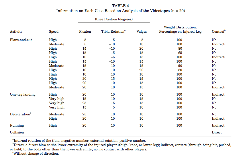
 Table from 'Injury mechanisms for anterior cruciate ligament injuries in team handball: a systematic video analysis' (Olsen 2001)

Kobayashi et al (2010) performed a twenty-year clinical research of 1,700 athletes in Japan. They found non-contact accounted for 60.8% of injuries with a knee in-toe out dynamic alignment in 51.9 % of cases. This reported dynamic alignment would likely be associated with abduction and externally rotation at the knee joint.

Boden (2000) found 72% of 100 ACL injuries were non-contact. The knee again was seen to be at near full extension. Mechanisms were sudden deceleration prior to a change of direction or landing motion with Valgus collapse of the knee.

Olsen also explored why the injury occurred even though the athlete may have performed this action many times. In many cases a perturbation was reported, as well as being pushed or held and having an unusually wide foot position. An ability of the motor system to deal with unexpected events is always a bonus and another variable we can factor into rehabilitation and prevention.

Here we see a reasonably consistent set of movement variables and positions that resulted in ACL injury. The point of having this data is that we must base our rehab and prevention strategies on the information we have available.

How does this guide us?

  • Knee flexion angle can be used to vary tension during rehabilitation.
  • Rehab & prevention using shallow knee flexion angles could be used as a high-level or late stage strategy.
  • Movement involving simultaneous multi plane kinematics and movements vital.
  • Whole leg sequencing of movement and force dissipation.
  • Velocity can be graded during rehab.
  • % on a single leg can be graded during rehab and used 100% during prevention.
  • Landing, deceleration & cutting motions vital for late stage and return to play.
  • Additional training variables such as perturbation or reactive elements are important.

This is a short video using some of the strategies discussed on a multi plane single leg squat task.

Hip, knee & ankle

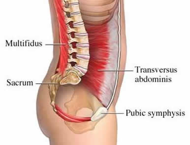
In any dynamic sport we involve the use of the whole kinetic chain to decelerate and summate force. With many ACL injuries we see a large amount of motion occurring from the knee in all three planes of movement.

We must also explore the ability of the hip and foot on the ipsilateral side to provide adequate motion to reduce the need for movement and force to localized at the knee joint. During a cutting movement with the foot firmly planted, the change of direction can be initiated proximally on the planted leg driven by the opposite leg in opposite direction. This concurrent external rotation and abduction of the hip may reduce the dynamic valgus and relative external rotation at the knee by way of the femur.

The ability of the ankle to evert and relatively abduct (motions associated with pronation) in a controlled manner may also control motion at the knee by way of the tibia. Internal rotation of the tibia could be caused by an inability of the foot to effectively control the rate or range of pronation. External rotation moment at the knee could be caused by the foot being unable to go into a pronated position reducing internal rotation of the tibia in close association with the femur.

Reduced ankle dorsi flexion and hip flexion could also reduce the amount of flexion the knee is able to go through leading to a shallow flexion angle associated with ACL injury.

Focusing on the skilled movement of the leg as a whole maybe a beneficial strategy to integrate the 3 components of the leg involved with planting and cutting and landing. The complex coordination and sequencing of multi joint and muscle actions is vital to the dissipation of the high forces that are required to cause the rupture of the ACL.

  • Skilled & sequenced movement of leg during function related movement.
  • Good active ROM of hip and ankle through all 3 planes for segmental contribution.
  • Focus on collective coordination & control of a specific movement rather than just the strength of individual muscles.

Here we see a number of points that can be taken from the research and our knowledge of the functional demands of sport to start to form specific rehabilitation and prevention programs. Research into more function related return to play tests that reflect the specific  movement demands of sport are needed, especially those that can be implemented without expensive clinical equipment.

In part two we will start to look at implementing the information we have gathered into a progressive rehabilitation program and the associated variables that can be manipulated.

References

Abrams G, Review Functional Performance Testing After Anterior Cruciate Ligament Reconstruction: A Systematic Review, The Orthopaedic Journal of Sports Medicine, 2(1) 2014

Boden BP, Dean GS, Feagin JA Jr, et al. Mechanisms of anterior cruciate ligament injury. Orthopedics. 2000;23:573-578.

Fayad L Anterior cruciate ligament volume: analysis of gender differences, J Magn Reson Imaging. 2008 Jan;27(1):218-23

Gottlob CA, Baker CL Jr, Pellissier JM, et al. Cost effectiveness of anterior cruciate ligament reconstruction in young adults. Clin Orthop Relat Res 1999;(367):272–82.

Hewett T et al, Biomechanical Measures of Neuromuscular Control and Valgus Loading of the Knee Predict Anterior Cruciate Ligament Injury Risk in Female Athletes - A Prospective Study, The American Journal of Sports Medicine, Vol. 33, No. 4 2005.

Kiapour A,Timing sequence of multi-planar knee kinematics revealed by physiologic cadaveric simulation of landing: Implications for ACL injury mechanism, Clinical Biomechanics 29 (2014) 75–82

Kobayashi et al, Mechanisms of the anterior cruciate ligament injury in sports activities: A twenty-year clinical research of 1,700 athletes, Journal of Sports Science and Medicine (2010) 9, 669-675

Markolf KL, Burchfield DM, Shapiro MM, et al. Combined knee loading states that generate high anterior cruciate ligament forces. J Orthop Res 1995;13:930–5.

Matsumoto H, Suda Y, Otani T, et al. Roles of the anterior cruciate ligament and medial collateral ligament in preventing valgus instability. J Orthop Sci 2001;6:28–32.

Mazzocca AD, Nissen CW, Geary M, et al. Valgus medial collateral ligament rupture causes concomitant loading and damage of the anterior cruciate ligament. J Knee Surg 2003;16:148–51.

Moseley L, Reconceptualising pain according to modern pain science, Physical Therapy Reviews 2007; 12: 169–178

Myklebust G,  Bahr R, Return to play guidelines after anterior cruciate ligament surgery, Br J Sports Med 2005 39: 127-131

Olsen, O.E., Myklebust, G., Engebretsen, L., Bahr, R., 2004. Injury mechanisms for anterior cruciate ligament injuries in team handball: a systematic video analysis. Am. J. Sports Med. 32, 1002–1012.


After another hugely enjoyable 'Functional therapeutic movement' course over the weekend we decided to pull together some of the key points from the course that form the basis of the Cor-Kinetic philosophy.


  • Functionality is about understanding the individual and their needs & how to bridge the gap between the two.
  • Sensitivity of an area is different to tissue damage. Being able to explain the peripheral physiology and neurobiology in simple terms will help!
  • You have to learn it first.
  • There maybe no relation between effectivness of therapeutic movement and increases in strength or ROM
  • Patients have different markers of success from therapists
  • Therapeutic movement aims:

Fearless

Freedom

Relaxation/flowing

Variation/variability

Meaningful/relevant

Explorative

  • Pain by definition involves the CNS
  • We can have 'optimal loading' but also 'tolerable' loading which is completely non functional but sometimes 'optimal'
  • Input does not always need to look like output.
  • There is no ‘right’ way to move. We can help people to move differently and provide different strategies.
  • Context is key.
  •  Motion is lotion.
  •  Movement is a whole body integrated experience.
  •  Movement can be used as a targeted input like any other input such as manual therapy.
  •  All movements are skills.
  •  Motor skills live in the brain.
  • We all have different structures, experiences and tolerances.
  • We need information from multiple assessments to form a movement strategy.
  •  The longer pain persists the weaker the relationship becomes with the state of the tissue. (The issue is not always in the tissue)
  •  Pain affects motor skills. Previous pain can affect current motor skills.
  •  Getting out of pain is often gold standard of treatment. Restoring a good level of movement ability is often not as important and sometimes ignored all together.
  •  Using movement as a treatment is simple. Pain free, free flowing, natural & varied rather than controlled and contrived.
  • Exercises are different to movement. Exercises tend to be controlled and contrived.
  •  Pain does not equal pathology in all cases.
  • Biomechanics still have a role to play in movement. 'Faulty' biomechanics maybe less relevant than previously thought.
  • Posture is only important if you can't move away from that one position. See it as a starting point - Don't assume it can't move.
  •  Give as few instructions as possible. Allow a large element of self organization.
  •  Practicing movement skills can alter our cortical representations and neurotags and also create new neurotags.
  • Pain is always an output of the brain.
  • Try not reinforce the pain experience or pain neurotag.
  • Immediate changes are secondary to long term changes.
  • Discomfort is different to pain. Some maybe be normal and should be experienced in a graded way.
  •  People in chronic pain can have altered movement skills and cortical representations.
  •  The brain pays attention to novel stimulus. High level of cortical excitement. Provide different and varied movement challenges.
  • Be a helpful passenger in the process not a driver. Give people tools to help themselves and not be reliant on a therapist.
  •  We do not ‘fix’ people but facilitate change and provide a nurturing experience for the individuals system to try and resolve the issue.
  • If your strategy is not working, try something else!
  • Know when to refer out to another skill set or discipline.
  •  Be careful with the language you use. It affects people negatively and positively.
  •  Movement affects nerves, skin, bones, muscles, connective tissues and the brain.
  •  If movement has no role in the problem then movement may not help.
  •  Restoring movement confidence can be key.
  •  Using a graded exposure to movement can be vital to movement change and toleration.
  • Increasing tissue toleration, desensitising people to movement and helping people to become more robust may help reduce future issues.
  • Build on pain free movement success.
  • Conscious & concentrated practice is needed to change subconscious movement behaviors.
  • Passive inputs may not change movement behaviours in the longer term. No associated learning process.
  • A positive outcome in a single session is secondary to long-term changes.
  • Helping people understand pain and specifically their interaction with it can be powerful.
  • Movement variables that can be manipulated.

Speed

Load

Range of movement

Local or global

Stability

Time

Frequency

Dimensionality

  • Many things can affect movement such as beliefs, stress, diet, mechanics, culture, disease and previous experiences.
  •  Provide a positive and stress free experience.

Want to know more about Functional Therapeutic Movement?

This is a webinar on the memory-prediction model of brain function and why it may guide some of our reasoning and techniques when it comes to assessment, treatment or training.

It is designed to be simple and easy to listen to rather then heavy on the neuroscience.

Below we have a rundown of the contents.


  • Memory - Prediction model. Is it important?
  • Bayesian Inference
  • The brain is not a computer. Its slow & predicts
  • Patterns, perception & prediction.
  • Auto association and recall of stored neural patterns.
  • Catching the ball
  • Hebb - "neurons that fire together. wire together"
  • Allport & inhibition
  • Neurotags, Moseley & Disinhibition.
  • Neurosignature, neuromatrix & Melzack
  • Processing of sensory information. Perception.
  • Importance of context
  • Visual, vestibular & proprioceptive.
  • Perception of threat & protective response.
  • Perception top down and bottom up
  • Top down altered perception of injured or painful area.
  • Processing of bottom up encoded signals.
  • Bayesien Inference - Prior knowledge & data = Beliefs
  • Forward model of motor control - D. Walpert
  • Predicting the feedback
  • Betting shop - Baysian process!
  • Stiffness/flexibility is often a prediction - Stepping off the curb.
  • Cortical hierarchy - Stored patterns.
  • How does this affect model influence our assessment & treatment or training?
  • Authenticity of movement or position to assess prediction output.
  • Pain as a prediction. Learned experience of pain.
  • Creating new memories & predictions. Hopefully positive!

There are many methods and treatments, especially relating to lower back pain, that include a focus on ‘core’ and ‘trunk’ stability and activation for the reduction of pain and increase in performance. In part one we will focus on the impact of a ‘core stability’ approach on chronic low back pain cLBP.

It would seem a common prescription for lower back pain would be a program of ‘core stabilization’ or ‘core strengthening’ both from healthcare professionals and the layperson alike. Its widespread clinical use (Xue-Qiang Wang et al 2012) as well as popular media coverage evidences this.


core.stability.muscles

Whilst there are varying powerful theories on activation patterns, implementation and importance of specific structures, this piece will focus on the actual efficacy of the approach by looking at the research and evidence into clinical outcomes such as pain, disability and fear avoidance beliefs.
The bottom line is this is what people really care about, simply if it gets rid of their pain or not. The efficacy of an approach to reduce pain can only be judged on its ability to do so.

By looking at a number of studies, especially systematic reviews or meta analyses this hopefully takes some of these variations into account between study parameters, methodology and sample size.

With such widespread use for cLBP there has to be something in it, doesn’t there?

A matter of timing


Hodges and Richardson carried out the original research into trunk muscle activation in LBP subjects in the mid & late 1990’s. Much of their focus was on the delay in timing of activation of Transversus abdominis in subjects with LBP. Their conclusion from “Inefficient muscular stabilization of the lumbar spine associated with low back pain. A motor control evaluation of transversus abdominis” was

“The delayed onset of contraction of transversus abdominis indicates a deficit of motor control and is hypothesized to result in inefficient muscular stabilization of the spine”

They studied TvA and trunk activation in a number of papers including “Delayed postural contraction of transversus abdominis in low back pain associated with movement of the lower limb” and “Altered trunk muscle recruitment in people with low back pain with upper limb movement at different speeds” with consistent delays in TvA and trunk muscle activation in anticipation of various limb movements recorded.

A lack of lumbar stabilisation was hypothesised to be a factor in pain experience of LBP patients, seemingly with a primary focus on the TvA. This has led to the implementation of exercises to stabilise the lumbar spine and improve the onset of activation of the TvA and other abdominal muscles to reduce ‘instability’ and pain.

One of the first questions we should ask, is the delayed timing a cause or effect of pain? If it is an effect then a focus on the activation may prove to be a fruitless endeavour. That is why we must also look to the research rather than theory as an indicator of efficacy. Are we seeing a correlation rather than causation?

The timing differences that existed between symptomatic and asymptomatic patients were about 20 ms, an exceptionally small measure of time, one fiftieth of a second. The measurements were, it is important to point out, all centred on timing rather than strength, although the terms ‘core strengthening’ has also gained popularity. To my knowledge there has been nothing to suggest increase in strength improves timing. Exercises that focus on conscious or volitional activation may be tough given the that such a small measure of time maybe beyond the conscious control of the patient.

It would also be worthwhile to get a measure of how the muscles of the trunk activate and there relative timings in different movement patterns so as build up a picture of the core's functional performance as well as clinical performance. This may give us a more rounded understanding of the core's activation.

Vasseljen et al (2012) in “Effect of core stability exercises on feed-forward activation of deep abdominal muscles in chronic low back pain: a randomized controlled trial” looked at the change in activation of trunk muscles over an 8 week period in response to a core stability program comprising low load core stability exercises or high load sling exercises. This was as well as a general exercise control group. They then compared feedforward trunk muscle activation to a similar arm movement as performed in Hodges original research.
After 8 weeks of this RCT involving a low load core stability group, a high load sling exercise group and a general exercise group, the onset of abdominal muscles had changed by between 15 and 19 ms only for the high load sling exercises compared to the core stability and general exercise groups.

No actual changes in pain were reported by any of the groups.

They concluded

“Abdominal muscle onset was largely unaffected by 8 weeks of exercises in chronic LBP patients. There was no association between change in onset and LBP”

Moreside et al (2013) also looked at trunk muscle activation in healthy and recovered lower back pain patients in their paper “Temporal patterns of the trunk muscles remain altered in a low back injured population despite subjective reports of recovery”

They found that although the recovered patients, who displayed perceived readiness to return to work and low pain scores, still had an altered muscle activation pattern and greater overall amplitudes of muscle activation.

In these cases it would appear that a restoration of ‘normal’ muscle activation timing and activity was not necessary for their pain experience to subside or caused the pain experience to subside. Equally it proved fruitless to attempt to change these activation patterns significantly with both high load and low load exercises. Any change recorded did not correlate with a reduction in pain.

It maybe suggested that these people were not actually free from the problem as they still displayed muscle activation changes. This cannot be ruled out. A follow up to see if pain and/or muscle activation changed in the future would shed more light on this. However  focusing on the timing of activation of the trunk did not seem particularly effective in the cases here in the time frames studied.

In normal day-to-day treatment clinicians do not have expensive EMG machinery to identify apparently ‘faulty’ firing patterns. The only measures that can be used are perceived readiness and pain scores.

Hodges, alongside Tucker, in their 2011 paper “Moving differently in pain” gives a different perspective to the activation of muscles in chronic pain patients.

“Existing theories predict relatively stereotypical change in whole-muscle behavior, but this has not been observed, and variable patterns of adaptation are identified in clinical populations”

He goes on to say

“changes in behavior of other muscles are unique to the individual and possibly to the task. This is most common in complex systems such as the trunk, where the muscle system has considerable redundancy (multiple muscles achieve a similar goal)”

The fact that we see no stereotypical change in muscle behavior should limit our attempt to create a consistent stereotypical behavior. How do we know what to change muscle behavior too? If multiple muscles achieve a similar goal then different people may use different muscular strategies to get the job done. There is a huge possibility that there is no ‘right’ way but many ways between the muscles associated within the task and varying from task to task, activation being function specific.

One of my favourite quotes is from famous British anatomist Charles Beevor in the 19th century. He simply stated in Beevors Axiom:

"The brain does not know muscles only movement"

He also gave a 1903 address to the royal college of physicians entitled "On Muscular Movements and their Representation in the Central Nervous System" This is simply way ahead of its time!

A redistribution of intra and inter muscular activity and mechanical behavior has been proposed to protect against pain and redistribute load. This could also happen when there is also the threat of pain. Inhibition and excitation in painful and surrounding areas/muscles has been clinically documented (Hodges 2011). The outcome goal of the activation pattern/timing would be to provide a protective strategy.
The nervous system may have a huge range of peripheral and central options to increase, decrease or redistribute activity. Such as motor neuron excitability, cortical inhibition or changes in motor planning.

An attempt to find a singular cure all for LBP seems to be a tall order with many factors across physical and psychosocial playing a part.

A Prospective study of back pain of 3,020 aircraft employees “A prospective study of work perceptions and psychosocial factors affecting the report of back injury” (1991) found that job satisfaction was a key factor in developing back pain. Subjects who ‘hardly ever” enjoyed their job were more 2.5 times more likely to report back injury than subjects who ‘almost always’ enjoyed their jobs. The researchers here took into account individual physical, psychosocial and workplace factors. An important point to note is that a history of current or recent back problems was also a factor in future back pain development.


Efficacy


Xue-Qiang Wang et al (2012) carried out “A Meta-Analysis of Core Stability Exercise versus General Exercise for Chronic Low Back Pain” The aim being to study the specific efficacy of a core stability approach. A Meta analysis provides more precision than a single study and also allows for variation in approaches and sample sizes of individual studies however, sufficient homogeneity is required. With many variations between approaches in the catchall term of “core stability” this study type is important.

The researchers here only included RCT’s (randomised control trials) that compared Core stability versus General exercise. They focused on pain intensity, back specific functional status, quality of life and work absenteeism. Out of 28 studies only 5 filled the research criteria. An RCT is regarded as the gold standard approach for determining the efficacy and effectiveness of an intervention in the hope of determining cause-effect relationship between treatment and outcome. Many studies into core stability fail to include all the necessary criteria, such as a control group, that allows us to make worthwhile associations of the intervention, taking out a third factor that maybe linked to intervention and outcome such as natural healing or regression to the mean which can be seen in cyclic pathologies such as CLBP. Hence the small number of included studies in this meta analysis. Sometimes we get a control group that also does nothing so essentially we are studying something against nothing which often favours the something group rather than comparing two differing interventions. However this decision is usually based on ethics. If evidence suggests usual care has no effect, then ethically a ‘nothing’ control group can be used. If however, evidence suggests whether usual care has an effect and the aim is to compare an intervention e.g. core stability exercises versus usual care, then usual care ethically should be given.

This threw up some interesting results. In the short term measures of pain and disability for the core stability intervention were better than for general exercise. However taking into account that we are discussing chronic back pain, no significant differences were observed at 6 months. So although providing some short-term effects, a core stability approach was an ineffective treatment of chronic lower back pain.
Their actual conclusion being

“Compared to general exercise, core stability exercise is more effective in decreasing pain and may improve physical function in patients with chronic LBP in the short term. However, no significant long-term differences in pain severity were observed between patients who engaged in core stability exercise versus those who engaged in general exercise”

One possible explanation for better short-term outcomes for the core stability group could relate to the lower load/intensity of some core stability exercises and therefore a better toleration from the patient. This however is pure hypothesis!

Mannion et al (2012) in “Spine stabilisation exercises in the treatment of chronic low back pain: a good clinical outcome is not associated with improved abdominal muscle function” looked at the link between increased ability to activate the transversus abdominis, obliquus internus and obliquus externus during "abdominal-hollowing" during rapid arm movement. Again this was consistent with the original research parameters from Hodges.
Although some improvements were made in disability and average pain levels pre to post therapy they did not significantly correlate to changes in muscle activation. Of the muscles tested only the TvA made any improvements in voluntary contraction, this being 4.5% (P=0.045).

They conclude that

“Neither baseline lateral abdominal muscle function nor its improvement after a programme of stabilisation exercises was a statistical predictor of a good clinical outcome. It is hence difficult to attribute the therapeutic result to any specific effects of the exercises on these trunk muscles”

Marshall et al (2013) looked at “Pilates exercise or stationary cycling for chronic nonspecific low back pain: does it matter? a randomized controlled trial with 6-month follow-up”
Again we see in this study that in the short term targeted specific lower back (SEG) exercises seem to have a better outcome than less targeted interventions. In this case cycling. Disability was significantly reduced in specific trunk exercise group after 8 weeks as was pain but to a lesser degree. Fear avoidance beliefs were reduced in the SEG group after 8 weeks compared to 6 months with stationary cycling group.

At 6 months however no difference between the two groups was reported with regards to clinically meaningful changes. As the patient group was of the chronic nature short term measures of success are less important than longer-term outcomes. In the longer term as the results for both interventions are similar the authors conclude that we could recommend either for chronic LBP.

Are the short-term outcomes for core stability interventions attributable solely to core stability or another mechanism such as patient beliefs? It would be useful to understand why the improvements are better in the short term versus the lack of long-term efficacy when compared against more general interventions.

Interestingly the researchers had this to say:

“The magnitude of improvement in self-report measures reported in the SEG may have been confounded by a number of participant biases toward receiving Pilates, a clinically more common type of exercise rehabilitation for LBP compared with stationary cycling”

Core stability is well marketed for cLBP whereas patients may perceive general movement to be damaging even though the long term outcomes are similar.

Conclusion


Although we see some short term benefits with ‘core stability’ exercises compared to more general exercises this approach does not seem to be more effective over longer time frames. Considering the needs of the patient group are of a chronic nature this type of intervention would have to be regarded as ineffective as a specific form of treatment in chronic patients. Could receiving a perceived clinically ‘relevant’ treatment influence short-term beliefs? Or perhaps better tolerated due to lower loads? It would be interesting to shed more light on the possible mechanisms behind this.

The proposal of a stereotypical adaptation to pain and, therefore intervention may not be a true reflection of the many and varied muscle activation adaptations to pain that are available to the CNS. The muscular activation pattern of the cLBP patient is likely to be individual and task dependent based on more modern EMG research findings into adaptations to pain (Hodges 2011).

Timing of muscle activation does not seem to positively or negatively correlate with reduction in pain levels or perceived readiness for return to work. Attempts to change timing of activation would appear to make little impact on actual onset of activation.
Approaches that combine a view of pathophysiological, motor control and psychosocial factors may stand the best chances of success, although a review of these approaches is the not the aim of this piece.

Many movement and motor control exercises maybe beneficial for cLBP. It is possible that no one form of intervention is right for all cases, especially in light of variable activation strategies and this seems to be highlighted by the research findings.

An increase in variation in available movements and specific movement strategies based around the individuals functional deficits and task specific needs may yield the best results with a movement based problem.Treating the individual would appear to be the only option if we can find no objective deficit to treat.

A generalised approach to exercise may be no better than an approach using ‘core stability’ exercises generically when a focus on individuality is used. As with most chronic injuries education about pain, movement and relationship to pathology alongside other applicable interventions maybe the best course of action.

Continuing education (CE) outside of a professional qualification is always going to be a good thing. The more we can learn and expose ourselves to information the better.  I am always reminded of a profound statement from a friend a few years ago in which he said:

“Have you got 10 years experience or 1 years experience, 10 times”

This can sometimes sum up some peoples evolution within their chosen profession.
Read more

This article is a standalone piece and also kind of serves as the third piece of 'The Brain, Movement and Pain' series already released earlier on this blog site.
Read more

The perspective that the body is an interconnected unit that displays regional interdependence is a valuable one. That different parts interact in different ways during different activities and influence ROM (range of movement) in other areas of the movement chain should seem a fairly easy link to make when looking at the whole body during different context dependent movements. We often eschew the value of the integrated system in favour of the isolated joint/muscle model.
Read more